Foot & Ankle
Our surgeons are often leading the way in foot and ankle care research. We are committed to advancing patient care through research, which results in advancements in the field and ultimately improves the care every patient receives.
The Achilles Tendon connects the gastrocnemius and calk muscles of the lower leg to the heel. It functions to pull the foot and ankle down during walking and running. An Achilles tendon rupture or tear occurs when the tendon is partially or completely torn due to being overstretched.
Causes
The area of the Achilles that is most frequently torn is near the heal. This area is vulnerable during activities that require lots of direct, forceful jumping and landing. Examples of such activities include basketball and gymnastics. Other potential causes of an Achilles tendon tear include falling and stepping from high to low surfaces (i.e. a hole). Males between the age of 30-40 who participate in recreational sports are most at risk to experience an Achilles tendon rupture.
Symptoms
The first and most noticeable sign of an Achilles tendon tear is a loud popping noise that heard immediately after the rupture. In some cases, the pop is loud enough to be heard by others near the patient. Other common symptoms include the following:
- Pain
- Swelling
- Inability to push off the foot
- Difficulty standing on the toes
Patients who experience the symptoms of an Achilles tendon rupture should make an appointment with an Orthopedic foot and ankle specialist. Diagnosing and treating the injury early is the key to treatment success.
Diagnosis
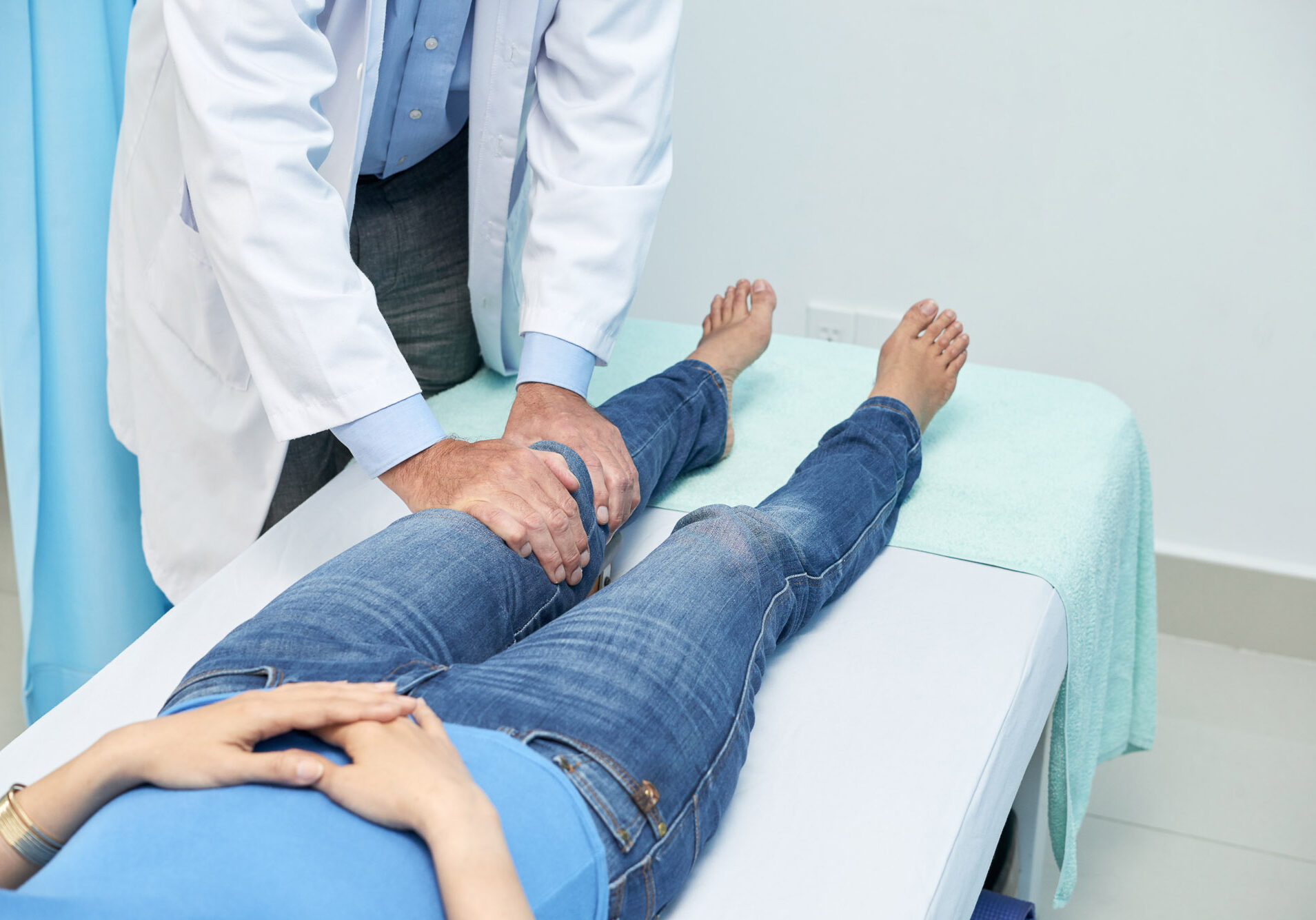
A physical examination is used to diagnosis an Achilles tendon rupture. During a thorough physical examination, the Orthopedic performs a series of tests to access the severity of the tear. An x-ray may need to be ordered to see if a piece of the heel bone came off during the rupture and an MRI may need to be done to confirm the Orthopedic’s diagnosis.
Treatment
Non-surgical treatment involves the use of a splint or cast. While the tendon is immobilized, it’s torn fibers heal.
Surgical treatment involves the use of sutures and special suturing techniques to repair the tendon and if necessary reattach it to the heel.
The Achilles tendon is the largest tendon in the body. Anatomically, it connects the gastrocnemius and soleus muscles of the calf to the heel. The tendon is responsible for key movements while walking, running, and jumping. Achilles tendonitis occurs when the tendons become irritated and inflamed due to overuse or injury. The two types of Achilles tendonitis are:
- Noninsertional Achilles tendonitis. The swelling and inflammation of tendon fibers located in the middle of the Achilles tendon.
- Insertional Achilles tendonitis. The swelling and inflammation of tendon fibers located near the insertion point of the Achilles tendon (the heel).
These may be caused by any of the following factors:
- Performing too much exercise or physical activity
- Starting a workout program before being physically ready
- Not stretching the calf muscles frequently
- A bone spur on the heel
Symptoms present to varying degrees based on the severity of the tendonitis.
Symptoms
The following symptoms may accompany Achilles tendonitis:
- Pain
- Stiffness
- A noticeable lump
- Swelling
Symptoms generally increase with weight bearing activities. When left untreated symptoms may become more frequent and severe.
Diagnosis
A medical history and thorough physical examination are used to diagnose Achilles tendonitis. Often times, medical imaging studies in the form of an x-ray or MRI may be ordered to ensure another type of injury is not causing the patient’s symptoms.
Treatment
Nonsurgical treatment options include rest, physical therapy, anti-inflammatory medications, and possibly immobilization. Braces and orthotic inserts may also be recommended. When the condition does not respond to these treatment options, a corticosteroid injection may be used to quickly improve symptoms and calm inflammation.
Surgical treatment is offered in two forms. The resecting of part of the gastrocnemius can help relieve the symptoms of some patients. In others, a debridement and tendon repair may produce better results.
The toe and forefoot are made up of 5 metatarsal and 14 phalange bones. Any of them can be fractured for a variety of reasons, the most common of which include the following:
- Foot to ground impact activities
- A direct blow
- A heavy object being dropped on the area
- Being step on
Fractures vary in the severity and complexity. A stress fracture is a fracture that occurs due to performing too much high impact activity. A nondisplaced fracture is one in which pieces of fractured bone maintain their alignment. A displaced fracture is one in which fractured bone pieces do not align properly. Open fractures are those that protrude through the skin. Special attention is shown to these types of fractures, especially in the foot because of infection risks.
Symptoms
Symptoms typically present immediately after the fracture. They usually include the following:
- Pain
- Swelling
- Bruising
Patients who suspect they have a fractured toe or forefoot bone should make an appointment with an Orthopedic foot and ankle specialist.
Diagnosing a Fracture
A physical exam and x-rays are used to diagnose toe and forefoot fractures.
Treatment
Toe fractures are treated by managing symptoms while the fracture heals. Anti-inflammatory medications help reduce pain and inflammation. Buddy taping a fractured toe to one that is not fractured help patients move their toe without experiencing increasing amounts of pain. A post-operative shoe is a flat sole shoe that evenly distributes weight across the foot.
Surgical treatment is typically reserved for severe fractures. Once the fracture is located, it is reduced and fixated using rods, plates, screws, and/or wires. It generally takes 6-8-weeks for a fractured bone to heal.
Special attention is drawn to fractures of the head of the 5th metatarsal (a Jones fracture). These fractures are easily fixed using a special compression screw. When tightened, a compression screw pulls pieces of fractured bone together.
Elevated blood glucose levels due to diabetes can cause nerve damage and decrease circulation. This can lead to problems for diabetics, particularly those that injure their foot. The nerves and blood vessels that supply the foot with sensation and blood are the first to be affected by nerve damage and circulation issues. Diabetics who wound or injure their foot may do so and not even know it. A fracture that is walked on will increase in severity and eventually deform the foot and/or ankle. When this happens, a condition known as diabetic or Charcot foot is said to have developed.
Symptoms
Key symptoms of Charcot foot that patients should look for are swelling, redness, and a visible deformity.
Diagnosis
A complete medical history, thorough physical examination, x-rays, and possibly a bone density scan are used to diagnose Charcot foot. Patients are also examined for wounds that they might not know about and that could potentially lead to infection.
Treatment
Nonsurgical treatment
Charcot foot cannot be cured. Treatment is focused toward managing symptoms and slowing the progression of the condition. Common nonsurgical treatment options include the following:
- Immobilization. Patients wear a cast, splint, or boot to help protect their foot and reduce swelling.
- Assistive devices. Crutches, canes, walkers, and wheelchairs can be used to help patients move around without putting weight on their foot.
- Diabetic shoes. Custom made shoes that fit perfectly and take pressure off the feet.
Treating Charcot foot is a team approach. The foot and ankle specialist and the patient work together to find a treatment solution that works.
Surgical Treatment
Severe fractures and dislocations due to Charcot foot may require surgical intervention. Because the procedure is difficult to perform and infection risks are high, most foot and ankle specialists try to avoid surgery as much as possible.
Plantar Fasciitis is a condition in which the tissue on the bottom of the foot becomes irritated and inflamed. Because the plantar fascia tissue supports the arch and is constantly under stress, managing plantar fasciitis can be difficult. Additionally, the symptoms of the condition can also be very uncomfortable for some patients, particularly those that like to constantly be on their feet. Luckily, there are numerous treatment options available for plantar fasciitis. When a foot and ankle specialist and patient work together to create a treatment plan tailored to their unique condition, maximum symptom relief occurs.
Causes
Plantar fasciitis results from overuse. Patients who are at the greatest risk to develop the condition fall into the following categories:
- Patients with a high arch
- Patients that are obese
- Patients that have tight calf muscles
- Competitive runners and walkers
The presence of a bone spur under the heel also puts patients at risk. Not all bone spurs under the heel are problematic. The ones that do, typically increase the severity of symptoms.
Symptoms
Symptoms are experienced under the foot and near the heel, they include pain and burning that typically gets worse with activity.
Diagnosis
A thorough physical examination is used to diagnose plantar fasciitis.
Treatment
Nonsurgical treatments
Nonsurgical treatment options include the following:
- Rest and ice. Slowing down or stopping activities that involve constant foot to ground impact or prolonged standing gives inflammation a chance to calm down. Icing painful areas serve the same purpose.
- Topical anti-inflammatory medications. When applied directly over symptomatic areas, these unique medications decrease inflammation and pain.
- Physical therapy. Stretching exercises loosen the tissue and calf muscles.
- Steroid injections. Powerful anti-inflammatory medications are injected directly over inflamed tissue.
- Shoes/inserts. Special shoes and inserts cushion the feet while walking and running.
- Splints. Keep the foot in a neutral position.
In most cases, a treatment program will include multiple treatment options. The overwhelming majority of plantar fasciitis cases will respond well to non-surgical treatment and not need to treated surgically.
Surgical Treatment
Two procedures may be used to treat plantar fasciitis. A gastrocnemius recession lengthens the calf muscles to improve flexibility and decrease tightness. During a plantar fascia release, the plantar fascia is partially cut and a bone spur is shaved down.
The Posterior Tibial Tendon stabilizes and supports the arch of the foot. When it is dysfunctional, patients experience pain. This condition is referred to as posterior tibial tendon dysfunction (PTTD). It is one of the most common conditions treated by a foot and ankle specialists.
Causes
PTTD is typically caused by inflammation that occurs due to injury or overuse. Patients who are most at risk as those that participate in sports that require lots of running and jumping.
Symptoms
The chief symptom of PTTD is pain along the tendon. Pain typically becomes worse with physical activity and prolonged standing.
Diagnosis
A medical history and thorough physical examination are used to diagnose PTTD. A foot and ankle specialist examines the patient and performs a variety of tests to determine exactly why the patient is experiencing symptoms. Once this is known, a treatment plan is developed.
Treatment
Numerous nonsurgical treatment options are available, including the following:
- Rest and ice. Slowing down or stopping activities that involve constant foot to ground impact or prolonged standing gives inflammation a chance to calm down. Icing painful areas serve the same purpose.
- Topical anti-inflammatory medications. Medications that can be rubbed over painful areas.
- Temporary immobilization. A cast, splint, or walking boot keeps the ankle immobilized so it can heal.
- Steroid injections. Powerful anti-inflammatory medications are injected directly over the inflamed tendon.
- Shoes/inserts. Special shoes and inserts cushion the feet and reduce pain.
- Night splints. Keep the foot in a neutral position.
- Physical therapy. Stretching exercises improve flexibility.
PTTD that is not improved with nonsurgical treatment options may require surgical intervention in the form of the following procedures:
A muscle recession or tendon lengthening. Surgical lengthening of the gastrocnemius muscle and Achilles tendon.
Tenosynovectomy. The inflamed tendon is surgical removed.
Tendon transfer. A tendon is taken from the foot and is used to replace the dysfunctional posterior tibial tendon.
Osteotomy. Bone is cut to reshape the arch of the foot.
The decision regarding which surgery to perform is based on the patient’s condition and the opinion of the foot and ankle specialist.
A bunion is a bony enlargement or prominence over the big toe. The majority of bunions do not need to be treated or are effectively treated using nonsurgical treatment options. Severe, painful bunions that alter the shape and function of the foot may require surgical intervention. The most commonly performed bunion surgeries include the following:
- Soft Tissue Repair. Tissues around the toe are tightened or loosened to help perfectly align the joint. Soft tissue repair is typically performed along with a bony procedure, such as an osteotomy.
- Osteotomy. Small cuts shape bone and realign the joint. Screws and metal plates are used to hold the bone and joint in place. Osteotomies are often performed in conjunction with a soft tissue procedure similar to the one described above.
- Arthrodesis. Severe bunions and severe arthritis are treated by removing arthritic bone and then fixating healthy bone using screws and plates.
- Exostectomy. Usually performed as part of a larger procedure, an exoctectomy is used to remove bony bumps over the joint.
- Resection Arthroplasty. A damaged joint is partially removed or resected.
Following a successful operation, joint anatomy and function are restored and pain decreases. The time frame for a full recovery is dependant upon the procedure performed. The decision regarding which procedure to perform depends on the patient’s condition and the option of the foot and ankle specialist treating it. Good candidates for bunion surgery are patients who have tried all other treatment options and have not found relief. It is important to note that foot and ankle Orthopedic specialists are the most qualified medical professional who performs bunion surgery.
Nerve damage and circulation problems associated with diabetes put diabetic patients at an increased risk to develop foot and ankle problems, including but not limited to Charcot foot and wounds. Diabetic patients should pay extra close attention to their feet and also take special care of them to ensure they remain healthy and functional. The following information can be used by diabetics or people who are taking care of a diabetic.
General care of the foot is important. Diabetics are encouraged to wash and dry their feet every morning—taking proper precautions to ensure the water is not too hot and the toes and feet are completely dry. Toenail trimming can be done at the diabetic foot clinic or at home. Special precautions should be taken when cutting. Nails should be cut straight across and corners should be avoided.
Inspection of the foot is also important and encouraged. The inspection should occur every day. It is important to check for blisters or cuts in between the toes and under the foot and heel. If abnormalities are found they should be taken care of right away. If an appointment is needed, it should be made.Shoes and orthotics should also be the ones that you and your foot and ankle specialist decided were best. They should always be kept clean and dry.
Other tips include:
- Avoid walking barefoot around the house
- Use lotion to prevent dry skin and flaking
- Avoid putting your feet directly in front of the air conditioner or heater
- Wear socks at night while in bed
Diabetics who have questions regarding how to take care of their feet or who have a current problem with their feet are advised to make an appointment with a foot and ankle specialist.
Our Specialties
The team at OSNI covers a wide variety of specialties for orthopedic needs. Take a look at them all below and reach out to our team by visiting our contact page to schedule a visit.